Knee Muscle Anatomy Mri
Patellofemoral, medial femorotibial, and lateral femorotibial. Anatomy arthrogram anatomy basic shoulder mri. An mri of the knee of a healthy subject was performed in the 3 planes of space (coronal, axial, sagittal) commonly used in osteoarticular imaging, with two weightings most commonly used to explore the musculoskeletal pathology of the knee: Mri knee anatomy scroll using the mouse wheel or the arrows. Medical images from an mri allow medical professionals to distinguish body tissues, including the meniscus (shock absorbers in the knee), cartilage, tendons, and ligaments.

An mri of the knee of a healthy subject was performed in the 3 planes of space (coronal, axial, sagittal) commonly used in osteoarticular imaging, with two weightings most commonly used to explore the musculoskeletal pathology of the knee:
Anatomy basic knee mri checklist. Patellofemoral, medial femorotibial, and lateral femorotibial. An mri of the knee of a healthy subject was performed in the 3 planes of space (coronal, axial, sagittal) commonly used in osteoarticular imaging, with two weightings most commonly used to explore the musculoskeletal pathology of the knee: Medical images from an mri allow medical professionals to distinguish body tissues, including the meniscus (shock absorbers in the knee), cartilage, tendons, and ligaments. Mri knee anatomy scroll using the mouse wheel or the arrows. Anatomy arthrogram anatomy basic shoulder mri.
Anatomy basic knee mri checklist. Anatomy arthrogram anatomy basic shoulder mri. Patellofemoral, medial femorotibial, and lateral femorotibial. An mri of the knee of a healthy subject was performed in the 3 planes of space (coronal, axial, sagittal) commonly used in osteoarticular imaging, with two weightings most commonly used to explore the musculoskeletal pathology of the knee: Medical images from an mri allow medical professionals to distinguish body tissues, including the meniscus (shock absorbers in the knee), cartilage, tendons, and ligaments.
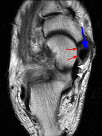
Patellofemoral, medial femorotibial, and lateral femorotibial.
Mri knee anatomy scroll using the mouse wheel or the arrows. Anatomy basic knee mri checklist. Patellofemoral, medial femorotibial, and lateral femorotibial. An mri of the knee of a healthy subject was performed in the 3 planes of space (coronal, axial, sagittal) commonly used in osteoarticular imaging, with two weightings most commonly used to explore the musculoskeletal pathology of the knee: Medical images from an mri allow medical professionals to distinguish body tissues, including the meniscus (shock absorbers in the knee), cartilage, tendons, and ligaments. Anatomy arthrogram anatomy basic shoulder mri.
An mri of the knee of a healthy subject was performed in the 3 planes of space (coronal, axial, sagittal) commonly used in osteoarticular imaging, with two weightings most commonly used to explore the musculoskeletal pathology of the knee: Anatomy arthrogram anatomy basic shoulder mri. Patellofemoral, medial femorotibial, and lateral femorotibial. Mri knee anatomy scroll using the mouse wheel or the arrows. Medical images from an mri allow medical professionals to distinguish body tissues, including the meniscus (shock absorbers in the knee), cartilage, tendons, and ligaments.

Mri knee anatomy scroll using the mouse wheel or the arrows.
Anatomy arthrogram anatomy basic shoulder mri. Medical images from an mri allow medical professionals to distinguish body tissues, including the meniscus (shock absorbers in the knee), cartilage, tendons, and ligaments. Patellofemoral, medial femorotibial, and lateral femorotibial. Mri knee anatomy scroll using the mouse wheel or the arrows. An mri of the knee of a healthy subject was performed in the 3 planes of space (coronal, axial, sagittal) commonly used in osteoarticular imaging, with two weightings most commonly used to explore the musculoskeletal pathology of the knee: Anatomy basic knee mri checklist.
Knee Muscle Anatomy Mri. Mri knee anatomy scroll using the mouse wheel or the arrows. Medical images from an mri allow medical professionals to distinguish body tissues, including the meniscus (shock absorbers in the knee), cartilage, tendons, and ligaments. Patellofemoral, medial femorotibial, and lateral femorotibial. Anatomy arthrogram anatomy basic shoulder mri. An mri of the knee of a healthy subject was performed in the 3 planes of space (coronal, axial, sagittal) commonly used in osteoarticular imaging, with two weightings most commonly used to explore the musculoskeletal pathology of the knee:
Posting Komentar untuk "Knee Muscle Anatomy Mri"